The
electron microscope
The electron microscope is based on the discovery by Thomson, Davisson and Germer that electrons have wave properties.
The wavelength, l needs to be of the same order as the size of an atom.
Example calculation:
If an atom has a spacing of 1.0 x 10⁻¹⁰m then calculate the anode voltage required:
atom has a spacing of 1 x 10⁻¹⁰m therefore:
l = 1 x 10⁻¹⁰m
mass of the electron, m = 9.1 x 10⁻³¹ kg
Planck constant, h = 6.6 x 10⁻³⁴ Js
charge on an electron, e = 1.6 x 10⁻¹⁹ C
square both sides of equation:
l2 = h2
2meV
rearrange to give V:
V = h2 = 149.6 Volts
2mel2
If an electron is accelerated through a p.d. of 500 000 V, then the wavelength associated with it is 1.73 x 10⁻³ nm. This means that if it can be used in a microscope the resolving power (which depends on wavelength) would be very large.
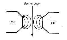
Electrons are also affected by magnetic fields, and magnetic focusing had been used for some time in the cathode ray tube. Then in 1926 it was realised that a suitable magnetic field arrangement could be used to act as a magnetic lens, bringing electrons of a given velocity to a focus and so giving an image of an object.
By combining several such magnetic lenses a succession of magnifications may be obtained, and this next diagram shows the similarity between an electron microscope and its optical counterpart.
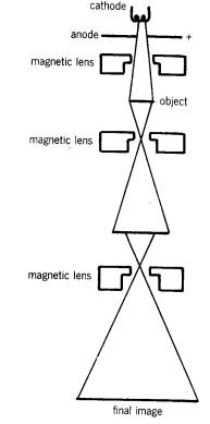
The condenser lens produces a parallel beam of electrons, which strike the object. Some electrons are absorbed by the object, some are transmitted and some are scattered sideways. These scattered electrons cannot pass through the small slit placed in front of the object, and in order to reduce the number of scattered electrons the object must be very thin.
The transmitted electrons pass through one or two magnifying lenses and the final image is formed on a screen or on a photographic plate.
The whole interior of the apparatus must be maintained at a very high vacuum, otherwise scattering of the electron beam from gas particles would ruin the image.
The magnification may be varied by altering the current in the magnetic lenses.
Accelerating voltages of 1 MV have been achieved, and for such an instrument the field in the lenses reaches a maximum of some 2.5 T using currents of 5 A in 3 000 turns, giving a focal length of about 5 mm. Such lenses must be cooled and work has been carried out on superconducting magnets for use in magnetic lenses. Theoretically the resolution of such an instrument would be very high, but aberration in the lenses limits it to about 0.1 mm. Magnifications of over 100 000 times are quite feasible, however.
Other problems exist at these very high magnifications: the objects that we are trying to view are little larger, by a factor of 1000 or so, than the electrons in the beam and so the electron beam distorts the object. Also the very high-energy electrons leak across the viewing screen, so blurring the image further.